")
Plant and animal cells and tissues (A-level biology)

Cells and tissues
Overview
The cell is the basic unit of life. A cell carries out a number of activities including protein synthesis, cell division, and is where hereditary materials are. Specialized cells constitute tissues.
General objective
By the end of the topic, the learner should be able to describe the structure, function, and organization of cells in an organism.
Specific objectives:
The student should be able to
- Identify plant and animal cell structures visible under the light microscope.
- Draw and label detailed animal and plant cells.
- State the functions of cell structures as seen in an electron microscope
- Distinguish the plant and animal cell as seen through an electron microscope
- Explain the theory behind the structure of the plasma membrane
- Describe the fluid mosaic model of the plasma membrane.
- There are two types of cells
- Prokaryotic cells have DNA that is not enclosed in a nuclear membrane. They do not have true nuclei. They lack membrane-bound organelles. Examples are bacteria and blue-green algae,
A diagram of a bacterial cell

2. Eukaryotic cells contain membrane-bound organelle including a true nucleus, mitochondria, chloroplast, and others.
Advantages of membrane-bound organelle
- Many metabolic processes involve enzymes embedded in membranes, so the membrane-bound organelles increase the surface area for activities like respiration and photosynthesis.
- Maintain enzymes of a particular pathway in one area for easy accessibility.
- The metabolic pathway inside an organelle can be controlled by controlling substances that enter and leave the organelle.
- Harmful reactants can be isolated
Differences between eukaryotic and prokaryotic cells
| Prokaryotic cell | Eukaryotic cell | |
| 1. | DNA not enclosed in a membrane | DNA enclosed in a double membrane nucleus |
| 2. | Has small ribosome (70s) | Has big ribosome (80s) |
| 3 | Lack mitochondria | Has mitochondria |
| 4 | Has no endoplasmic reticulum | Has endoplasmic reticulum |
| 5 | Small size | 1000-1000 times the size of prokaryotes |
| 6 | Lack of lysosomes and peroxisomes | contain lysosomes and peroxisomes |
| 7 | Lack of Golgi apparatus | Has the Golgi apparatus |
| 8 | Simple flagella | Complex flagella |
| 9 | Respiration occurs in mesosome | Respiration occurs in mitochondria |
| 10 | Lack chloroplasts | Plant cells have chloroplasts |
| 11 | Have the ability to fix nitrogen | Lack of ability to fix nitrogen |
Fine structure of an animal cell

Fine structure of plant cell

Differences between plant cell and animal cell
| Plant Cell | Animal cells | |
| 1 | Has chloroplast | Lack chloroplast |
| 2 | Has a cell wall | Lack of cell walls |
| 3 | Has large central vacuole | Lack a vacuole or has small vacuoles |
| 4 | Has starch granule | Lack of starch granule |
| 5 | Nucleus at the side | Nucleus centrally placed |
| 6 | Tonoplast present around the vacuole | Tonoplast absent |
| 7 | Centrioles absent | Centrioles present |
| 8 | Cilia and flagella absent in higher plants | Cilia and flagella present |
| 9 | Few cells are capable of division | Almost all cells are capable of division |
Parts of the cell
Cytoplasm
All cells have a cytoplasmic matrix. It is an aqueous solution or colloidal suspension of mainly vital cellular materials. It is the site for protein synthesis and metabolic activities
Functions of the cytoplasm
- Contain or keep organelles
- Contains nutrients for organelles
- Stores materials
- Maintain conducive atmosphere for a cellular reaction
Cell membrane
It serves as a boundary between the cell and its environment. It may permanently exclude some items from the cell while permanently retaining others.
Functions of the cell membranes
- It separates the contents of the cell from the external environment.
- Controls exchange of materials between the cells and external environment
- It separates compartment with specialized functions inside the cell
- Acts as a receptor site for recognizing external stimuli such as hormones.
- Allows uptake of materials by phagocytosis and pinocytosis.
- Support enzymes of complex metabolic pathways in place for close proximity.
The fluid mosaic model of the cell
- The cell membrane is about 7nm thick
- It consists of a continuous phospholipid bilayer in which proteins are scattered in a mosaic manner.
- Proteins penetrate through the bilayer at a varying degree to form intrinsic and extrinsic proteins
- Intrinsic proteins are those that penetrate through the bilayer while extrinsic proteins are those that float on top as islands in a sea.

Components of the cell membrane and their function
- Phospholipids: affect the fluidity and permeability of the membrane
- Cholesterol: make the membrane less fluid at higher temperatures.
- Glucolipids: act as recognition sites e.g. human blood group system is a result of different glycolipids on the cell membrane of red blood cells.
- Proteins:
- provide structural support for the membrane,
- assist in active transport across the membrane
- act as recognition sites
- act as enzyme, energy transducers and electron carriers
- Glycoproteins are recognition sites, e.g., for neurotransmitters and hormones.
The nucleus
This is the largest cell organelle enclosed by a double membrane perforated by nuclear pores. It contains chromatin which is the form of chromosomes during interphase. The nucleus also contains nucleolus that produces ribosomes
Function of nucleus
- Contains DNA for inheritance controlling cell division and protein synthesis.
- The nucleolus manufactures ribosome
- Controls all activities of the cell
28. Growth in size of a single cell is limited by the
A. cytoplasm.
B. nucleus.
C. cell vacuole.
D. cell membrane.
Nuscloli
Functions of nucleoli
The nucleolus is the site of synthesis of ribosomal RNA which is combined with proteins in the cytoplasm to make ribosomes.
Mitochondria
It a cell organelle surrounded by two membranes, the inner being folded to form cristae. The mitochondrion contains a matrix with a few ribosomes a circular DNA molecule and phosphate granules. Its main function is producing energy by aerobic respiration.
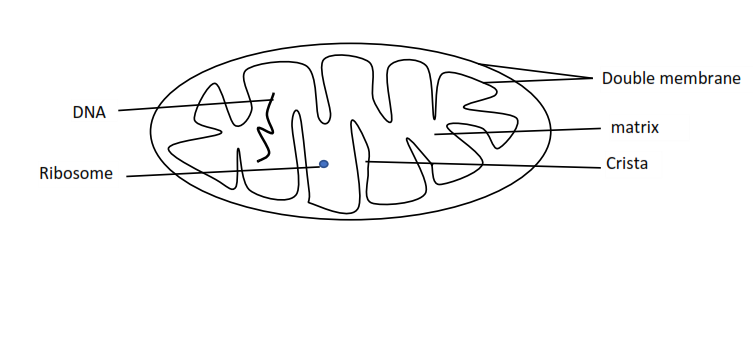
Adaptations of mitochondria to its function
- The inner membrane is folded to form cristae that increase surface area for enzymatic activities.
- Contains circular DNA to produce the necessary enzymes.
- Has a large surface area for diffusion of gases.
- Matrix contain necessary enzymes for Krebs cycle
Endoplasmic reticulum
This is a system of flattened membranes bound sacs called cisternae, forming tubes and sheet. Is continuous with the outer membrane of the nuclear envelope. Some of its parts is covered by ribosome and this is called rough endoplasmic reticulum. The part without ribosomes is called smooth endoplasmic reticulum.
Functions of endoplasmic reticulum
- Ribosomes are site of protein synthesis
- Smooth endoplasmic reticulum is a site of lipids and steroid synthesis.
- The tubes are for intracellular transport
Golgi apparatus
Consists of stack flattened membrane-bound sacs, called cisternae, continuously being formed at one end of the stack and budded off as vesicles at the other.
Functions of Golgi apparatus
- Producing glycoproteins by adding carbohydrates to proteins
- Producing secretory enzymes, e.g. digestive enzymes
- Replenishing the cell wall
- Produces materials for synthesis of plant cell wall.
- Produces lysosomes concerned with breakdown of worn out structures in the cell.
Lysosome
Single small organelle that contain enzymes to destroy worn out parts of the cell and autolysis (digestion of the cell)
Chloroplasts

It is restricted to plant cell and used for photosynthesis. It is surrounded by an envelope of two membranes and contains a gel-like stroma through which runs a system of membranes that are stacked in places to form grana. The stroma contains ribosomes, circular DNS and lipid droplets.
Similarities between mitochondria and chloroplasts
- Both have double membrane, circular DNA
- Inner membrane is folded to increase the surface area
- Contain ATPase enzyme for ATP synthesis
- Both occur in plant
- Both contain carrier proteins
- Both contain circular DNA
- Both contain ribosome,
Differences between mitochondria and chloroplasts
| Chloroplast | Mitochondria |
| Structural difference | |
| Contain chlorophyll | Does not contain chlorophyll |
| Inner membrane form grana | Inner membrane folded to form cristae |
| May contain starch granules | Does not contain starch granules |
| Functional difference | |
| Use water | Produce water |
| Produce O2 | Produce CO2 |
| Use sunlight and store its energy in food made | Set energy free from food for work |
| Occur only in green plants | Occur in both plants and animals |
Histology
Specific objective
The learner must be able to
- Explain how epithelial tissues are adapted to the diversity of functions in the body
- Distinguish between different levels of the organization
- State the advantages and disadvantages of being unicellular
- State the advantages of being multicellular
Definition
A tissue is a group of similar cells linked with associated intercellular substances to perform a particular function(s). In a complex organism, different tissues combine to form organs, and organs combine to form organ systems. Organ system combine to form the organism
| Advantages of unicellular state | Advantages multicellular state |
| Can exist on its own | There is specialization |
| Do not need gaseous exchange surface | Indefinite growth |
Types of animal tissues
Classification of tissues depending on their function leads to the following:
Animal tissues
- Epithelial tissues
- Connective tissue
- Skeletal tissue
- Nerve tissue
- Reproductive tissue
Plant tissue
- Meristematic tissue
- Epidermal tissue
- Parenchyma
- Collenchyma
- Sclerenchyma
- Vascular
- Cork
Animal tissues
Epithelium
These are tissues that cover the external and internal surfaces of the animal body. They may be made up of one or more layers of cells resting on a basement membrane. The cells are connected together by a substance called hyaluronic acid.
The epithelial tissues function to protect underlying structures from injury through abrasion or pressure and from infection. Stress is combated by the tissues becoming thickened and keratinized, and where cells are sloughed off due to contact friction the epithelium shows a very rapid rate of cell division so that lost cells are speedily replaced. The free surface of the epithelium often is highly differentiated and may be absorptive or secretory in function
Epithelial tissues are subdivided into two major categories
- Simple epithelium
- Compound epithelium
Simple epithelium
This type of epithelium is made up of only one layer of cells. Simple epithelium is divided into 5 types
- Squamous
- Cuboidal
- Columnar
- Ciliated
- Pseudostratified
Squamous epithelium
consists of a sheet of flattened cells which fit closely together rather like crazy paving

Location of squamous epithelial tissue
- Skin outer layer
- Bowman’s capsule in the kidney
- Alveoli of lungs
- Capillary walls
Squamous epithelium is thin and therefore allows easy diffusion of materials across it.
Function
- Protective
- Allow easy diffusion
Cuboidal epithelium
Heights of the cell are approximately equal to its width, when viewed in vertical section the cells appear square.

Location
- Salivary duct
- Collecting duct of the kidney
- Thyroid gland
Functions
- Secretory
- Absorptive and its surface may be increased by microvilli to increase surface area.
Columnar epithelium
It is made of elongated cells at right angles to the basement membrane.

Location
The lining of the stomach and small intestines
Functions
- Secretory e.g. secretion of mucus in the stomach
- Absorptive e.g. absorption of digested food in the intestines.
Ciliated epithelium

This is made of columnar shaped cells but having numerous cilia at their end. The cells are associated with mucus-secreting goblet cells producing fluids in which cilia set up the current.
Location
- Oviduct
- Trachea
- Bronchi
Pseudo-stratified epithelium
This is made up of one layer of cells but some cells do not reach the free surface. It appears as if it is stratified.

Location
- Urinary tract
- Trachea
- Olfactory mucosa
Function
Secretory
Compound epithelium
made up of more than one layer of cells. There are two types of compound epithelium
Stratified epithelium
Made up of a number of layers of cells. The cells are made by mitotic division of the germinal layer which rests on the basement membrane
Occurrence: vagina, esophagus, and skin
Function: protects the body against friction.
Transitional epithelium
This is made of 3-4 layers of the cell. The cells are able to modify their shape when placed under different conditions.
Location: urinary bladder, ureter, and pelvis
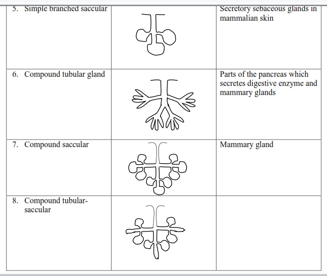
Glandular epithelium
These are epithelium cells that are folded inwards forming invagination where cells lining the bottom of the invagination are secretory.
There are two types
- Exocrine glands: these are glands whose secretion are released through ducts
- Endocrine glands: are glands without ducts and pass their secretion into blood streams.
Types of exocrine glands
There are different types of glands depending on the epithelial folding.


Connective tissue
These are the tissues that hold specialized tissues and organs in the right position and fill the spaces between them. They consist of jelly-like ground substances or matrix in which several types of cells are embedded.
Classification
- Loose connective tissue
- Fibrous tissue (white and yellow)
- Adipose tissue
- Dentine tissue
- Skeletal tissue
- Blood cell making tissue
Areolar
This is the fundamental type of connective tissue found allover the body beneath the skin and therefore connecting the skin and therefore connecting the skin to structures below it. It binds sheets of epithelium to mesenteric (capillary network around alimentary canal). It joins blood vessel and nerves whereby they enter or leave body organs. It also fills up space between adjacent tissue and therefore acts like packing tissue.
Functions
- packing tissue
- insulator due to accumulation of fat cells
- support other organs
Structure

Functions of parts of connective tissue
- Fat cells store fats
- Collagen and elastic fibers provide mechanical support and flexibility.
- Matrix provides nutrients to the cell
- Fibroblast produce the ground substance
- Neutrophil, macrophages, mast cells for defense
White fibrous tissue
This a tough tissue composed of an organized bundle of collagen fiber closely packed together and running parallel to each other. Rows of fibroblasts are scattered among the collagen and run alongside the bundles. Each bundle is bound to a neighboring tissue by areolar tissue. Fibrous tissue is abundant in tendons, ligament, sclera, and cornea of the eye. These are areas where great strength and limited flexibility is required.

Yellow fibrous tissue
This contains a glycoprotein matrix containing only elastic fibers. The fibers are irregularly arranged and are branched. Fibroblasts are randomly distributed throughout the matrix. The elastic fiber provides the tissue with elasticity and flexibility. It also contains some few bundles of collagen which give it strength. It is found in ligaments, walls of arteries as components of lung and associated passage.

Adipose tissue
This tissue has no specific matrix but closely packed fat-filled cells arrange in two lobules
The bone
Is a tissue that provided skeletal network in the body
Functions of bones
- It provides a shape that allows easy movement and recognition.
- Protect delicate parts of the body; for, example rib cage protects the heart and the lung
- Provide support
- Provide a means of attachment of the muscle to allow movement
- Store minerals like calcium and phosphorus
- Produce blood cells like a red blood cell
Structure
The bone is made of a matrix and cells.
The matrix of compact bone is made of collagen fibers together with inorganic substances such as calcium, magnesium, and phosphorous. These components are arranged in concentric circles called lamellae, around a Haversian canal containing an artery, a vein, lymph vessel, and nerve fibers.
Bone cells are found in spaces in the lamellae known as lacunae and fine channels called canaliculi link lacunae.
The system of lamellae around one Haversian canal is called a Haversian system.


Bone formation, also called ossification, is a process by which new bone is produced. Ossification begins about the third month of fetal life in humans and is completed by late adolescence. The process takes two general forms, one for compact bone which makes up roughly 80 percent of the skeleton, and the other for cancellous bone, including parts of the skull, the shoulder blades, and the ends of the long bones.
A bone of the first type begins in the embryonic skeleton with a cartilage model, which is gradually replaced by bone. Specialized connective tissue cells called osteoblasts secrete a matrix material called osteoid, a gelatinous substance made up of collagen, a fibrous protein, and mucopolysaccharide, an organic glue.
Soon after the osteoid is laid down, inorganic salts are deposited in it to form the hardened material recognized as a mineralized bone. The cartilage cells die out and are replaced by osteoblasts clustered in ossification centers. Bone formation proceeds outward from these centers.
This replacement of cartilage by bone is known as endochondral ossification. Most short bones have a single ossification center near the middle of the bone; long bones of the arms and legs typically have three, one at the center of the bone and one at each end. Ossification of long bones proceeds until only a thin strip of cartilage remains at either end; this cartilage, called the epiphyseal plate, persists until the bone reaches its full adult length and is then replaced with bone.
The flat bones of the skull are not pre-formed in cartilage-like compact bone but begin as fibrous membranes consisting largely of collagen and blood vessels. Osteoblasts secrete the osteoid into this membrane to form a sponge-like network of bony processes called trabeculae. The new bone formation radiates outward from ossification centers in the membrane. This process is called intermembranous ossification. There are several ossification centers in the skull. At birth, bone formation is incomplete, and soft spots can be felt between these centers. The lines where the new bone from adjacent centers meets form cranial sutures visible on the surface of the adult skull.
Both endochondral and intermembranous ossification produce immature bone, which undergoes a process of bone resorption and deposition called bone remodeling to produce mature bone.
Plant tissues
Simple plant tissues consist of only one type of cell. They are grouped according to the degree of thickening present in the cell wall
Parenchyma
It is a simple permanent tissue of unspecialized usually spherical cells with thin cell walls. Parenchyma form the bulky of packing tissue within the plant.

Functions of parenchyma tissue
- Store water and food reserve
- When tightly packed and turgid provide support for herbaceous plants
- It is a ground tissue
- Air spaces allow buoyancy in floating plants
- Air spaces allow gaseous exchange
Example
Which one of the following plant tissues performs both storage and supportive functions?
A. Parenchyma.
B. Sclerenchyma.
C. Collenchyma.
D. Phloem.
The answer is A
Parenchyma is the plant tissue that has both storage and supportive function. It store water and starch in most plants and also serves as the main supporting tissue in non-woody plants.
Collenchyma
It contains cells with additional cellulose deposited in the corners.

Function
- It provides mechanical strength to the petiole, leaves, and stem of young dicot plants.
- Collenchyma confers flexibility to various parts of the plant like petiole and stem, allowing for easy bending without breakage.
- It allows for the growth and elongation of plant organs.
- Collenchyma present in leaves also prevents them from tearing.
- The living cells of collenchyma store food.
- Collenchyma when containing chlorophyll performs the function of photosynthesis.
Sclerenchyma
Mature sclerenchyma cells are dead and cannot grow. They develop fully when the growth of the surrounding tissue is complete. Sclerenchyma cells have large deposits of lignin in the cell wall and the cell content is lost in places, lignin is not deposited due to the presence of plasmodesmata in the primary cell wall, such regions are called pits. Some sclerenchyma cells are roughly spherical and are known as sclereids. These are usually found in small groups in fruits and seeds, cortex, pith, and phloem.

Function of sclerenchyma
- They provide mechanical support
- They make up xylem and tracheid for water transport
- In hypodermis of xerophytic plant, they prevent water lost
- Sclerenchyma cells in the fruit walls help in its dehiscence and seed dispersal
- Sclerenchyma of seed coat protects the seed from desiccation
Xylem Consist of parenchyma cells and fibers together with vessels and tracheid.
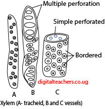
Vessels are made of cylindrical dead cells, one on top of another with the cross wall broken down to form a long continuous tube from the roots to the leaves.
The type of vessel found to depend on the degree and nature of cell thickening. In the protoxylem the lignin is deposited in rings or spirals to the cells is still capable of expansion. In metaxylem, there is more extensive lignification arranged in patterns known as reticulate, scalariform, or pitted.

Tracheids are spindle-shaped cells arranged in rows with ends of the cells overlapping. The cells have heavily lignified cell wall with no cell contents.
Functions of xylem
Transport water and mineral salts
They provide mechanical support.
Adaptations of the xylem
- Cross walls are perforated or completely removed to form continuous tubes from roots to stems and leaves
- Xylem vessels have no living contents to allow water to flow freely
- Contain bordered pits to allow water cross to live cells
- Lignified to prevent water loss
- Lignified to prevent them from collapsing under negative pressure of transpiration pull.
- Small tube to enable high capillarity
- Xylem walls have high adhesive forces.
Adaptation to provide support
- Walls are lignified
- Vessels are circular for additional support.
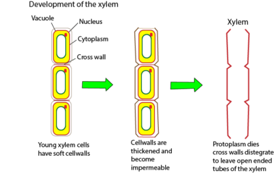
Development of xylem
Cells destined to form xylem vessels elongate and develop thickened secondary walls. The walls are later lignified. The cell content die and cross-section walls degenerate to form a continuous open tube.

Phloem


The phloem consists of sieve tubes and companion cells.
Sieve tubes consist of columns of elongated, thin-walled living cells called sieve tubes/elements. They have cross walls with many holes or pores called sieve plates. Each sieve tube has a companion cell.
Function
Transport of manufactured food (sucrose and amino acid) from leaves to other parts of the plant.
Adaptations
- Lack of a nucleus and most cell organelles to leave room for transportation of food
- The sieve plates are perforated to allow rapid flow through
- Has filament for quick transport by streaming
- has an intimate association with companion cells to obtain energy and materials
Differences between xylem and phloem
| Xylem | Phloem | |
| 1 | Vessels are made of dead cells | Elements are made from living cells |
| 2 | Vessels have lignified cell walls | Phloem do not have lignified cell walls |
| 3. | The end wall disappears completely | The end wall form sieve plates. They do not disappear completely |
| 4. | Have pits | Have plasmodesmata |
| 5. | Thick walls | Thin walls |
| 6 | Transport water and mineral salts | Transport food (sucrose and amino acids) |
Development of phloem
Cells destined to become sieve elements elongate, most cell organelles degenerate leaving cytoplasmic filament. The plasmodesmata of the end wall widen forming sieve pores.
For revision questions and answer download PDF
Please find free downloadable notes, exams and marking guides of agriculture, biology, and chemistry from digitalteachers.co.ug website.
Dr. Bbosa Science
The work is good and exceptional could you please include work on micro bodies ..peroxisomes and glyoxysomes
Ribosomes
Cilia and flagella
Cytoskeleton
The Role of the Acidity of N Heteroaryl Sulfonamides as Inhibitors of Bcl 2 Family Protein Protein Interactions, ACS Med can you buy cheap cytotec for sale While many groups around the world have been testing existing drugs for efficacy against COVID- 19 using cultured cells, it is well known that cells grown in a dish do not behave like the cells in a living human body, and many drugs that appear effective in lab studies do not work in patients