
Coordination, nervous system, hormonal system

Coordination and control in animals
Coordination means to cause the part to function together or in proper order.
Coordination and control in animals are performed by the nervous system and the endocrine system.
Difference between the nervous and endocrine system
| Nervous system | Endocrine system | |
| 1. | Fast acting | slow acting |
| 2. | It’s effects are localized | It’s effects are diffuse |
| 3. | Transmission is electrical and Chemical theory cell fibres | Relies on chemical transmission through the circulatory system |
| 4. | Transmission occurs in nerves | It occurs in a blood |
The nervous system.
The nervous system is composed of highly differentiated cells called nerve cells or neurons. Those that carry impulses from receptors to the control nervous system are called sensory neurons while those that carry impulses from the CNS to effector are called effector neurons.
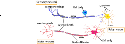
Functions of parts of nerve cell
- Nerve fibre or axon transmits impulses
- The myelin sheath protects the axon; it also insulates the axon and speeds up transmission of impulse.
- Nucleus controls cellular activity
- Dendrites or make contacts with other nerve cells or effector
- Nodes of Ranvier are microscopic gaps found within myelinated axons. Their function is to speed up propagation of action potentials along the axon via saltatory conduction
- The cell body preserves the structural integrity of the neuron, houses the genetic material, and supplies energy to drive activities.
Differences between effector and sensory neuron
| Effector neurons | Sensory neuron | |
| 1. | Transmit impulse from CNS to the effector | Transmit impulse from sense organ or receptor to the CNS cell body in the middle of axon |
| 2. | Cell body at the end of the axon | Cell body in the middle of the axon |
| 3. | Cell body located in the grey matter of the spinal cord | Cell body located in dorsal root ganglion of the spinal cord |
Structure of the nerve cell
- The cytoplasm contains the same organelles as the other body cell; mitochondria, nucleus cell membrane, and ribosome grouped in Nissl’s granules.
- A long nerve fiber, axon extend from the cell body in effector neurons or either side of the cell body in sensory neurons to transmits impulses. The axon enclosed within a fatty myelin sheath which is not part of the neuron but another cell Schwann cell which wraps itself repeatedly around the axon. The myelin sheath protects the axon but also insulates the axon and speeds up the transmission of the impulse.
Transmission of impulse.
The resting potential of axon
A potential difference is maintained between the inside and outside of the undisturbed axon. The inside being negatively charged [about- 70 ml] with respect to the outside. In this state, the membrane is said to be polarized. The resting membrane potential is maintained by the sodium/potassium pump using energy derived from ATP and it pumps ions against their concentration gradient pumping sodium ions are pumped out of the neuron in exchange for potassium ion (K+). K+ ions and organic ions is higher inside the neuron whereas Na+ and Cl– ion concentration is higher outside the neuron
The impulse
An impulse or action potential is a temporary and local reversal of the resting membrane potential, arising when an axon is stimulated. The term used during the electrical change which occur during the passage of action potential is depolarization, and axon is said to be depolarized. The action potential is short- lived lasting about a millisecond, after which the resting membrane potential is restored. Events occurring into axon during the transmission of action potential are show in the figure below.

Information is transmitted through the nervous system as a series of impulse which travels as an action potential.
Properties of nerves and impulses
Stimulation
In normal circumstance impulse are set up in nerve cell as a result of excitation of the receptor. But an impulse can be set up in nerve by applying any stimulus which opens the sodium channels and cause depolarization of the membrane. In general, nerves can be stimulated by mechanical, osmotic, chemical, thermal and electrical stimuli.
All or nothing law
An excitable tissue will only be excited by a stimulus above a certain threshold stimulus intensity. For any given neuron the amplitude of the action potential is always constant and increasing the strength or number of stimuli has no effect on this. For this reason, potentials are described as all or nothing event. The all-or-nothing law states that the response of an excitable unit (axon) is independent of the intensity.
Refractory period.
After an axon has transmitted an impulse, it cannot transmit another one straight away. The axon has to be recovered first. The membrane has to be repolarized and first.
The period under which the nerve cannot transmit an impulse following the transmission of the first one is called refractor period and typically lasts about 3 milliseconds. It can be divided into the absolute refractory period during which the axon is totally incapable of transmitting an impulse, followed by a somewhat longer relative refractory period during which it’s possible to generate an impulse in the axon provided that the stimulus is stronger than usual.
The importance of the refractory period, together with transmission speed, it determines the maximum frequency at which the axons can transmit impulses. For most axons the maximum frequency is about 500 per second, though some neurons can reach 1000 per second.
Factors that affect nerve conduction velocity
- Axon diameter
- Impulses move faster in an axon with larger diameter because longitudinal resistance of exoplasm decreases with increasing diameter of axon, which increases the length of the membrane influenced by local circuits as the distance between adjacent depolarization increases; causing increased conduction velocity.
- Small cells or cells with large surface area: volume ratio or ion leakage weakens membrane.
- Myelin sheath stops ion leakage; therefore, large diameter only important for unmyelinated neurons
2. Myelination and saltatory conduction:
- Myelination speeds up conduction. In a myelinated neuron, the conduction velocity is directly proportional to the fiber diameter.
- Schwann cells prevent diffusion of ions; flow of current occurs only between adjacent nodes of Ranvier
- Therefore, depolarization only at nodes of Ranvier because action potential jumps from node to node
3. Temperature: Homoiotherms with steady body temperature have faster impulse propagation than poikilotherms which have fluctuating body temperature.
4. Resting membrane potential (RMP): Effect of RMP changes on conduction velocity is quite variable. Usually, any change in the RMP in either direction (hyperpolarization or depolarization) slows down the conduction velocity.
The Synapse.
This a functional area, where an axon come into contact with another for the purpose of transferring information. These are two types of the synapse, electrical and chemical, depending on the nature of the transfer of information across the synapse, A structurally dissimilar but functionally similar synapse exists between the terminal of a motor neuron and the surface of muscle fibre and this is called the neuromuscular junction.
Structure of the chemical synapse
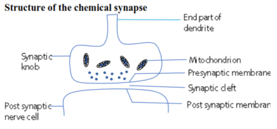
Transmission across a synapse.
- The arrival of an impulse at the synaptic causes an influx of Ca2+ ions into the knob from the synaptic cleft.
- The Ca2+ ion causes the synaptic vesicles to move towards the pre-synaptic membrane.
- The vesicles fuse with the presynaptic membrane and release a transmitter substance into the synaptic cleft by exocytosis.
- The transmitter substance diffuses across the synaptic cleft and attaches to specific receptor sites on the postsynaptic membrane.
- This causes an influx of Na+ ion into the postsynaptic membrane, resulting in local depolarization of the membrane. If the Na+ ion surge is large enough, an action potential (impulse) is generated in the postsynaptic neuron.
The transmitter substance.
The transmitter substance at the majority of the synapse is acetylcholine an ammonium base that has an effect on the permeability of the nerve membrane. After its effect, it’s inactivated by an enzyme cholinesterase to enable successive impulse to be kept separate. The products of this hydrolysis pass back into the synaptic knob where they are resynthesized into Acetylcholine using energy from ATP.
The Nerve- muscle junction or Neuromuscular.

Transmission across the neural muscular junction is the same as in any other chemical synapse. When an impulse arrives at the nerve muscular junction, acetylcholine is discharged from synaptic vesicle into the synaptic cleft. The acetylcholine diffuses across the gap and depolarizes the muscle endplate.
The action of drugs and poisons.
Chemicals that destroy acetylcholine, inhibit its formation, or prevents its action, will stop synaptic transmission.
Atropine, for example, doesn’t prevent acetylcholine being formed but it stops it depolarizing the postsynaptic membrane and therefore causes a synaptic block.
Curare, the poison used on the tips of arrows by south American Indians has a similar effect, especially on a nerve-muscle junction.
On the other hand chemicals (e.g. serine) nerve gases that destroy or prevent the formation or action of cholinesterase will be expect to enhance and prolong the effects of acetylcholine.
Strychnine also enhance synaptic transmission to such an extent that a person suffering from strychnine poisoning will give convulsive muscular contraction upon slightest stimulation.
Noradrenaline
This another neural transmitter substance in sympathetic nervous system of vertebrates. Nerve that produce acetylcholine are called cholinergic nerve and those producing noradrenaline are called adrenergic.
The amount of transmitter substances released by a synapse steadily falls off in the response to constant stimulation until the supply of transmitter substance is exhausted and the synapse is described as fatigued. Further information passing along this pathway is inhibited and the adaptive significance of fatigue in the prevention of damage effect due to overstimulation.
A post synaptic neuron receives stimuli from a variety of source and integrates them and produce a coordinated response.
Temporal summation at synapse enable weak background stimuli to be filtered out before it reaches the brain.eg information from the receptor in the skin, the eyes and ears receive constant stimuli from the environment which has little immediate importance for the nervous system. Only changes in the intensity of stimuli are significant to the nervous system and these increase the frequency of stimuli and pass across the synapse and evoke a response.
The transmission of information across a synapse and neuromuscular function may be prevented post-synaptically by the activity of certain chemical blocking agents or pre-synaptically.
The vertebrate nervous system.
The nervous system of vertebrates is characterized by the structural and functional diversity of neurons and their complex organization with the body. The nervous system is subdivided into two main parts, the central nervous system [CNS] and peripheral nervous system.
The central nervous system consists of the brain and spinal cord while the peripheral nervous system consists of numerous nerves that link the CNS with the receptors and effectors.
Reflex action and reflex arch.
The reflex action in a rapid, automatic stereotyped response to a stimulus that is not under the conscious control of the brain. It’s also described as involuntary action. The neurons forming the pathway taken by the nerve impulse in reflex action is referred to as reflex arch. Illustrated below.
The reflex arc

Conditioned reflex arch
This is a form of reflex action where the response is modified by the past experience, it’s coordinated by the brain. Learning for the basis of all conditioned reflexes such as salivation at sight or smell of food.
The autonomic nervous system
The autonomic nervous system is the part of the peripheral nervous controlling activities of the internal environment that normally involuntary such as heart rate, peristalsis, and sweating.
The autonomic system is divided into two parts;
The sympathetic and parasympathetic system. Both contain nerve fibers serving structures over which the body has little or no voluntary control. In both cases nerve fiber emerge from the brain or spinal cord and pass to the organs concerned. There are many pathways in each of the two systems. Along the course of each pathway, there is a complex set of synapses constituting a ganglion. The nerve fibers on the proximal side of the ganglion are called preganglionic fiber, those on the distal side postganglionic fibers,
The main structural difference between the parasympathetic and sympathetic system relates
the position the ganglion and are given below.
| Sympathetic | Parasympathetic | |
| Structural differences | ||
| 1. | ganglia lie close to the vertebrate | Ganglia are embedded in the walls of the effector |
| 2. | Preganglionic fibre short | Long preganglionic fibre |
| 3. | Long post ganglionic fibre | Short post ganglionic fibre |
| Functional differences | ||
| 4 | Transmitter is noradrenaline | Transmitter substance is acetylcholine |
| 5. | Accelerates heartbeat | Slow heartbeat |
| 6. | Dilate bronchioles | Constricts bronchioles |
| 7. | Dilates iris | Constricts iris |
| 8. | Slows gut | Speeds up gut movement |
| 9. | Constricts bladder and anal sphincter | Relaxes bladder and anal sphincter |
| 10 | Cause relaxation of the bladder | Causes contraction of the bladder |
| 11. | Contracts erector pili muscles | – |
| 12. | Increase sweat secretion | – |
| 13. | – | Stimulates tear glands |
| 14 | – | Causes flow of saliva and other secretion |
The brain
The brain is swollen anterior end of the vertebrate neural tube which has the over role of the coordination and control of the activities of the whole nervous system. To accomplish this there are special centers or nuclei in the different parts of the brain for dealing with specific functions such as locomotion, balancing and so on
Functions of the brain
1.Receives impulses from receptors
2.Integrates these impulses
3.sends out new impulses to the appropriate effect.

The functions of the main parts of the human brain.
- Medulla oblongata is the most posterior part of the brain. It contains centers controlling breathing, circulation, swallowing salivation and vomiting.
- The cerebellum coordinates voluntary movements such as posture, balance, coordination, and speech, resulting in smooth and balanced muscular activity. It is also important for learning motor behaviors
- The thalamus and associated structures: contain centers controlling such function as sleep, aggression, feeding, drinking, osmoregulation, temperature regulation, speech, and sexual activity.
- The pituitary is an endocrine gland that secretes a wide range of hormones controlling such function as water and salt balance, growth, metabolism, and sexual development.
- The cerebral hemispheres coordinate all voluntary responses.
Hormonal communication.
Hormones are organic compounds produced in one part of the body, from which is transported -usually in the bloodstream – to another part when it evokes a response.
In the human and other vertebrate hormones are secreted into the bloodstream by endocrine glands.
Position of the main endocrine gland in a human body
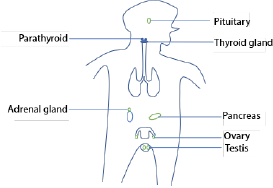
The principal endocrine glands of mammal, hormones and functions of the hormones
| Gland | Hormone | main function | |
| a. | Thyroid | Thyroxine | Raise the basal metabolic rate |
| Calcitonin | Opposes action of parathormone | ||
| b. | Parathyroid | Parathormone | Controls the concentration of calcium and phosphate ions in the blood |
| c. | Pancreas | insulin | lows blood sugar concentration |
| d. | Adrenal medulla | adrenaline | prepare the body for emergency; metabolic rate increases, |
| e. | Adrenal cortex | Aldosterone | Controls concentration of K+ and Na+ in blood |
| Cortisol | Prevent excessive immune response | ||
| androgens | Promotes the development of testes and secondary sexual characteristics | ||
| F | Pineal body | melatonin | causes concentration of melanin in frog’s skin; promote sexual development in mammal |
| g. | Testes | Androgens | Promotes development of testes and secondary sexual characteristics |
| h. | Ovaries | Estrogens | promotes development of ovaries secondary sexual characteristic of female control menstrual cycle and pregnancy. |
| I | Pituitary (anterior lobe) | Thyroid-stimulating hormone | Causes the thyroid gland to secrete thyroxine |
| Adreno -corticotrophin (ACTH) | Cause adrenal cortex to secrete adrenal cortical hormones | ||
| Growth hormones | Stimulate growth | ||
| prolactin | Causes mammary gland to secrete milk | ||
| Follicle-stimulating hormone | Controls testes and ovary | ||
| Luteinizing hormone | Controls testes and ovaries | ||
| Pituitary (posterior lobe) | Antidiuretic hormone (ADH) | Causes reabsorption of water in kidney | |
| oxytocin | Causes contraction of uterus at birth |
How hormones control cells
When a hormone molecule reaches a target cell, it binds to the plasm membrane at a specific receptor site. The receptor site is located on the outer surface of the membrane and is associated with a molecule of adenylate cyclase. The binding of the hormone to the membrane increases the activity of adenylate cyclase, causing ATP to convert into cyclic AMP. The cyclic AMP [Adenosine monophosphate] then activates enzymes that bring about the appropriate response within the cell. The extent of the particular response is determined by the concentration of cyclic AMP which in turn depends on a delicate balance between adenylate cyclase responsible for synthesis, and another enzyme phosphodiesterase – which destroys it.
For sample questions and sample, answers download the pdf below
Sponsored by The Science Foundation College + 256 753 802709
Compiled Dr. Bbosa Science
Thank you for the work you are providing to us as students of Uganda and i would like to request you that am an A level biology student offering BCM and have tried to search notes of coordination and i have failed to download them so i kindly request you to send me those
notes via my email
thank you
may God bless you
on digitalteachers.co.ug, click “science” the top bar menu,
Thanks for the updates but as a slow learner i would like to understand the work clearly plz,can u make it specific!
You have a real talent for this field. Home & Kitchen
Thanks for making this so enjoyable. Sports News
Explore the detailed MBBS Fees Structure in Karnataka, covering government and private institutions.
Secure direct access to premier institutions with MBBS Direct Admission in Rajasthan.
Use the Raja Luck Invite Code to get exclusive rewards and bonuses.
Get unique discount rates and cashback deals when you utilize the 82 Lottery Invite Code throughout registration.