")
Transport in animals (A-level biology)

Transport in animals
Transport is the movement of materials from one part of the organism to another. Transport involves diffusion and active transport in simple and small animals and at a cellular level.
Big animals require a mass flow circulatory system to deliver food materials and other essentials and remove waste products from the cell. This is because big animals have small surface area to volume ratio that they cannot meet their transport requirement by diffusion.
Advantages for circulatory systems in big animals
- Supplies metabolites and removes waste products from the cells at a faster rate than diffusion would do.
- It enables separation of materials transported; e.g. oxygenated blood is transported different vessels from those that transport deoxygenated blood.
- Impermeability of external surface to remove water. Example of the thick cuticle of insects.
- Avoids the utilization of materials along the way.
Development of transport system in Animals
In the course of evolution, advanced organisms attained a more advanced transport system than a primitive one. This can be shown in the following examples:
- Protozoans: transport their materials by cytoplasmic streaming. Simple diffusion across the membrane surface, facilitated diffusion and active transport as well as pinocytosis and phagocytosis.
- Cnidarians: transport by the movement of body wall to create water current in their body cavity, which circulates food, water and dissolved gases.
- Platyhelminthes: have very thin flattened shape enabling materials to be exchanged between the organism and the environment by diffusion.
- Annelids have a coelom separating body wall from internal organs (gut) so needs a system between the two regions to enable food, gases and waste products to be transported between the regions. An earthworm has a closed blood circulatory system with pigmented blood.
- Arthropods: have a hard exoskeleton so cannot depend on simple diffusion for the transport of materials between its tissues and the environment. They use a tracheal system to transport oxygen and carbon dioxide and have a hemocoel containing colorless blood in which organs are suspended. Thus, the body organs are in direct contact with blood. The blood does not transport oxygen and therefore does not contain hemoglobin.
- Vertebrates: circulatory system has muscular heat to pump blood through blood vessels throughout the body.
Features of circulatory systems
- Transport medium or blood to carry dissolved materials such as food oxygen and carbon dioxide.
- Vessels to carry the medium to all parts of the body
- A pump to propel the medium in the vessel
- One- way values to keep the medium flowing in one direction
- A close association between the tissue and medium so that the cell can obtain the required substances from the medium and deliver their waste products to it.
-
Transport medium
A few animals have a type of blood, which is more or less like seawater. One of the substances to be transported is oxygen. To increase oxygen-carrying capacity of water most active animals have pigments in their blood. In most species, the pigment is contained in special cell but in some species is free in the fluid part of blood the plasma.
Each pigment consists of a prosthetic group and a protein.
Comparison of oxygen-carrying pigments
| Pigment | Metal in the prosthetic group | Color of pigment | Example of animals | |
| Hemoglobin | Iron | red | vertebrate | Plasma or cells |
| Hemocyanin | Copper | blue | arthropods | plasma |
| Chlorocruorin | iron | green | annelid | plasma |
| Hemerythrin | iron | violet-pink | sipunculids, priapulids, brachiopods | Cells or plasma |
Structure of hemoglobin
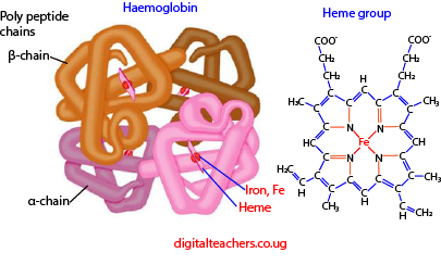
Structure of hemoglobin
- Hemoglobin comprises four subunits, each having one polypeptide chain and one heme group.
- The polypeptide chains of adult hemoglobin are of two kinds, similar in length but differing in amino acid sequence.
- The two alpha chains each has 141 amino acid residue while the two beta chains each has 146 amino acid residues
- Heme, which accounts for only 4 percent of the weight of the molecule, is composed of a ring-like organic compound known as a porphyrin to which iron in oxidation state II is attached.
- Oxygen binds reversibly to the ferrous iron atom in each heme group.
- The binding of oxygen to the heme group of one subunit has an effect of increasing the affinity of a neighboring subunit (on the same molecule) for oxygen.
Functions of hemoglobin
- The main function of hemoglobin is to carry oxygen from the lungs to all the tissues of the body.
- Some of the carbon dioxide is transported from tissues to lungs through hemoglobin. Although the majority of it is transported via plasma still it carries some of CO2 to lungs.
- Buffering action:
Hemoglobin also acts as a buffer. Buffer is a substance that resists change in pH. Blood has pH of 7.4 and it remains in the narrow range, because, if it changes, the life of the person may be endangered.
- Interaction with drugs: For not only oxygen but also hemoglobin act a very important role in the transport of various drugs to their site of action.
The blood
Blood is a specialized tissue consisting of several types of cells suspended in a fluid medium called plasma.
Functions of mammalian blood
1. Transport of soluble organic compounds from the small intestine to various parts of the body.
2. Transport of soluble excretory matters to organs of excretion.
3. Transport of hormones from glands where they are formed to target organs.
4. Distribution of heat in order to maintain body temperature
5. Defense against diseases, which may be obtained through blood clotting, phagocytosis, and immunity.
6. Maintenance of a right blood solute potential as a result of plasma protein activity.
7. Transportation of respiratory gases i.e., CO2 &O2
Components of blood
- Water:
This maintains blood pressure and volume. It’s where dissolved materials are transported around the body. The pressure is particularly important since the glomerulus requires high pressure to form urine.
2. Plasma proteins:
These include
(i) Serum albumen; abundant to increase the viscosity of blood and binds with calcium. Calcium is important for the functioning of enzymes.
(ii) Serum globulin which includes
- α-globulin, which binds with and transports hormone thyroxine, lipids, and fat-soluble vitamins; A, D, E, K.
- β-globulin, binds and transport iron, cholesterol, and fat-soluble vitamins; A, D, E, K.
- γ-globulin are antibodies produced by lymphocytes for immune response.
(iii) Prothrombin– a catalytic agent involved in blood clotting.
(iv) Fibrinogen- a protein involved in blood clotting.
(v) Enzymes- that control rate of metabolic activities in blood.
(vi) Mineral salts: include Na+, K+, Ca2+, Mg2+, HPO42-, HCO3–, Cl–, etc. they regulate osmotic pressure and pH level of blood. Ca2+ helps nervous transmission and blood clotting.
(vii) Dissolved products od digestion, excretory products, vitamins and hormones that are transported in the body
(a) Erythrocytes:
They produced in the liver in infants, embryo, and cartilaginous organisms or in bone marrow in those organisms that have bones. Their function is to carry oxygen.
Adaptations of red blood cells to its function
1. They have a biconcave disc shape to increase the surface area for the absorption of oxygen.
2. They lack a nucleus, which permits hemoglobin to be packed into the cell.
3. They are small therefore able to squeeze between capillaries
4. They have a thin membrane permitting efficient diffusion of gases (short distance for diffusion)
5. They contain hemoglobin, which has a high affinity for oxygen.
6. They do not carry out any metabolism so they do not utilize the oxygen being transported.
Carriage of oxygen
Oxygen diffuses into the red blood cells across its plasma membrane and combines with the hemoglobin to form oxyhemoglobin. The attachment of oxygen does not involve chemical oxidation of iron which remains in Iron (II) state throughout the process. The union is a loose one, the oxygen molecule being attached to hemoglobin in the lungs and equally readily detached in the tissues.
The efficiency of hemoglobin as an oxygen carrier
When one of the four polypeptide chains in the hemoglobin molecule receives oxygen molecule in the lungs its structure is altered in such a way that the remaining three polypeptide chains accept oxygen readily. In the tissue, the reverse occurs: one of the polypeptide chains loses its oxygen molecule and this causes the others to give up their oxygen more readily. In other words, hemoglobin takes up oxygen more rapidly if it already possesses one or more oxygen molecules, and releases it more rapidly if it has already released one or more oxygen molecules.
Oxygen dissociation curves
The ability of blood to transport enough oxygen to meet the needs of the body is largely attributed to the affinity of hemoglobin for oxygen.
This can be determined by exposing a sample of blood to different partial pressures of oxygen (concentration of oxygen in the air), and then determining the percentage saturation of blood with oxygen in each case.
A plot of percentage saturation of hemoglobin with oxygen against partial pressure of oxygen gives an oxygen dissociation curve below

The oxygen dissociation curve is S-shaped (sigmoid) showing that it is very appropriate for a blood pigment. Over the steeply rising part of the curve, a small increase in the partial pressure of oxygen achieves a relatively high percentage saturation of the blood.
The flat part of the curve at the top corresponds to the situation in the lungs: over this range, a high saturation is maintained even if the partial pressure of oxygen in the alveoli falls.
The oxygen dissociation curve favors the loading of oxygen in the lungs and offloading of oxygen in the tissue.
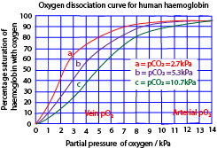
The graphs above show that a high concentration of carbon dioxide shifts the curve to the right. i.e. it lowers the affinity of hemoglobin for oxygen. The release of oxygen is therefore favored in the tissue where the partial pressure of carbon dioxide tends naturally to be high as result of its continual release from respiring cells. The oxygen uptake occurs in the lungs where the partial pressure of carbon dioxide is low.
In summary, the affinity of hemoglobin for oxygen is reduced by
- Low oxygen concentration.
- High carbon dioxide concentration
- High body temperature
- Low PH
High carbon dioxide concentration (or low PH) has the effect of establishing deoxyhemoglobin.
This favors the unloading of hemoglobin hence reducing its affinity for oxygen.

The oxygen dissociation curve of myoglobin lies at the left of that of hemoglobin because myoglobin has a high affinity for oxygen. Myoglobin is found in muscles; it picks oxygen from hemoglobin and stores oxygen to be used when the partial pressure of oxygen falls to a very low level as in severe muscle exertion.
Similarly, the hemoglobin of animals like lungworm, which burrows in oxygen-deficient mud, has a higher affinity for oxygen than hemoglobin.
The location of the oxygen dissociation curve of an organism to the left of another usually indicates a higher affinity for oxygen by its hemoglobin. Thus, the mountain gorilla has a high affinity for oxygen than many other animals. This allows its hemoglobin to get saturated at low oxygen tension. This enables it to survive in conditions of low oxygen partial pressure at high altitudes
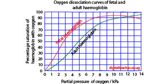
The oxygen dissociation curve of fetal hemoglobin lies at the left of that of adult hemoglobin; this because fetal hemoglobin has a high affinity for oxygen than adult hemoglobin this enables fetal hemoglobin to pick oxygen from the mother’s blood.
Effect of carbon monoxide on oxygen carriage of hemoglobin
Carbon monoxide combines more readily with hemoglobin than oxygen. This prevents hemoglobin to combine with oxygen, thus carbon monoxide is powerful respiratory poison.
Transport of carbon dioxide
CO2 reacts with water to produce carbonic acid H2CO3. In the red blood cells, the reaction is catalyzed by carbonic anhydrase.
Carbonic acid dissociates into hydrogen ions. Hydrogen ions combine with hemoglobin to form haemoglobinic acid while HCO3– diffuses into the blood.
The electroneutrality in the red blood cells is maintained by an inward movement of chloride ions from the plasma so-called chloride shift.
In the lungs, hydrogen ions combine with hydrogen carbonate ions to give CO2 which is lost to the atmosphere.
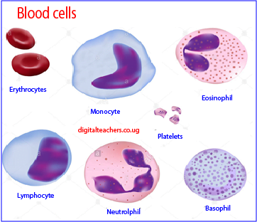
(b) White blood cells
These are produced in the bone marrow and are of various types:
(i) Granulocyte; this constitutes 72% of the total leucocytes. They have an irregular lobed nucleus and a cytoplasm containing granules. The granulocytes are divide into 3 types.
- The granules of neutrophils remain unstained when a dye, eosin is applied to them
- Eosinophils: stain red with the eosin dye. They are one of the immune system components responsible for combating multicellular parasites and certain infections in vertebrates. They possess antihistamine properties, which help to reverse allergic conditions. E.g. asthma or hay fever
Basophils. These stain blue with eosin dye. The cells produce heparin an anticlotting hormone and histamine in inflammation.
(ii) Agranulocytes;
These have a rounded or bean-shaped nucleus. Their cytoplasm has no granules They include lymphocytes and monocytes.
- Lymphocytes
These have a rounded nucleus with no cytoplasmic granules. They originate from the lymph nodes and cause antibody production and immediate immune reactions such as willing of tumors, grafts etc.
- Monocytes
These are formed in the bone marrow and have a bean-shaped nucleus. They are phagocytes and ingest bacteria
- Platelets; are produced in the bone marrow and initiate blood clotting.
These are irregularly shaped membrane-bound cell fragments frequently nucleated and made from the bone marrow. They are used in blood clotting.
Vascular systems in Animals
There two types of vascular systems: open and closed vascular systems.
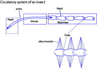
(a) The open vascular system: (most arthropods, some cephalad molluscs, tunicates).
Blood is pumped by heart into an aorta which branches into a number of arteries. These open into a series of blood spaces collectively called hemocoel. Here cells are in contact with the blood and materials are exchanged by direct diffusion through the plasma membrane.
Blood under low pressure, blood moves slowly between tissues gradually percolating back into the heart via open-ended veins. The distribution of blood in tissue is poorly controlled. This limits the efficiency of the open system.
Fortunately, gaseous exchange in insects takes place through the tracheal system. The insect circulatory system is not therefore concerned with transporting oxygen and carbon dioxide. Accordingly, it lacks oxygen-carrying pigment. However, it plays an important role in distributing food substances and eliminating nitrogenous waste.
(b) The closed vascular system: (echinoderm, cephalo, molluscs, annelids, vertebrate).
Here blood is confined in a series of specific vessels and not permitted to touch the body tissues. In animals with a closed system, the heart more muscular heart and blood are pumped by the heart rapidly around the body under sustained high pressure and back to the heart. Material exchange occurs across the wall of blood capillaries, which ramify through the organs and come into close association will all cells. Animals with closed systems are generally larger and often more active.
The disadvantage of the closed circulatory system is that the blood is contained in vessels whose walls form a barrier between the blood and the surrounding tissues. Oxygen and other materials have to cross this barrier. However, capillaries having very thin walls reduce this barrier.
(i) The single closed circulatory system
Here blood possesses through the heart once in every circuit of blood.
In fish, for example, blood is pumped from the heart to the gills. After acquiring oxygen from the gills, blood flows to the body tissues and then back to the heart.
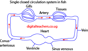
The problem with this arrangement is that blood has to pass through two capillary systems, the capillaries of the gills and then those of the body, before returning to the heart. Capillaries offer considerable resistance to the flow of blood, and this means that in fishes there is a marked drop in blood pressure before the blood completes a circuit. For this reason, the blood flow from tissues to the heart is sluggish. This is overcome to some extent by the fact that fishes have large sinuses, which offer minimum resistance to blood flow, in places of veins. Nevertheless, the problem of getting blood back to the heart is an acute one and probably imposes a severe limitation on the activities of many species of fish.
(b) Double Circulation
In double circulation, blood passes through the heart twice in every complete circuit.
Advantage
- Blood is pumped to the lungs at a much lower pressure than that at which it is pumped to the rest of the body. In humans, the pressure in the pulmonary artery is about one-sixth of that in the aorta.
- Deoxygenated blood is separated from oxygenated blood and then pumped at a different pressure to the lung and the body respectively.
(i) Incomplete double circulation
A frog’s heart has two atria and one ventricle, however, blood mixing in the ventricle is prevented by the spiral valve in the conus arteriosus a heart chamber immediately before blood is pumped into big arteries.
(ii) Separate hearts
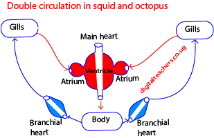
The squids and octopuses solved the pressure problem by having two hearts: blood is pumped from the main heart to various parts of the body. It flows through a system of sinuses to a pair of branchial hearts that pumps it to the gills.
The Human circulatory
The circulatory system consists of three independent systems that work together: the heart (cardiovascular), lungs (pulmonary), and arteries, veins, coronary and portal vessels (systemic). The system is responsible for the flow of blood, nutrients, oxygen and other gases, and as well as hormones to and from cell
The figure below shows the main blood vessel in the human Circulatory system

Arties and vein
Arteries transport blood at high pressure to the body while veins transport blood at low pressure from the body to the heart.
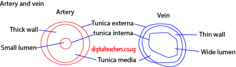
Adaptation of the artery
-thick wall to accommodate high pressure
– have narrow lumens to maintain high pressures
– some arteries like aorta valves to prevent the backflow of blood.
Adaptation of veins
-wide lumen to lower resistance to blood flow
-valves allow blood tom flow in one direction
Differences between arteries and veins
| Arteries | veins | |
| 1 | Thick wall | Thin walls |
| 2. | Narrow lumen | Broad lumen |
| 3. | Have no valves except pulmonary artery and aorta | Have valves |
| 4. | Carry oxygenated blood except for pulmonary artery | Carry deoxygenated blood except |
| 5. | Pulse detectable | Pulse not detectable |
| 6. | Empty at time of death | Get filled up at death. |
Capillaries
Is where exchange believe blood and cell takes place
Adaptation of capillaries
-thin walls for fast diffusion
The heart
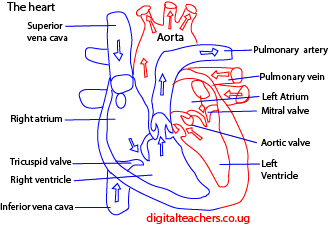
Blood returning via the vena cava enters the right atrium. The resulting pressure in this chamber forces and opens the flaps of the tricuspid valve. The result is that blood flows through the atrioventricular opening into the right ventricle.
When the atrium and ventricle are full of blood the atrium suddenly contracts, propelling the remaining blood into the ventricle. The contraction spreads from the right atrium over the rest of the heart. Atrial systole is relatively weak but the ventricles, whose thick walls are particularly well endowed with muscles, contract more powerfully. As a result, blood is forced from the right ventricle into the pulmonary artery.
The blood is prevented from flowing back into the atrium by the flaps of the atrioventricular opening. The atrioventricular valve is prevented from turning inside out by tough strands of connective tissue, the tendinous cord or “heartstrings” which run from the underside of each flag to the wall of the ventricle
Once in the pulmonary artery, blood is prevented from flowing back into the ventricle by pocket like semilunar valves guarding the opening of the pulmonary artery.
From the lungs, oxygenated blood returns to the left atrium via the pulmonary veins. It is then conveyed to the left ventricle and so into the systemic arch which leads to the aorta. The flow of blood takes place in the atrioventricular valve consists of two flaps rather than three, for which reason. It is called the bicuspid valve. It is also known as the mitral valve because its two flaps are rather like a bishop’s miter.
Although systole starts at the right atrium, it quickly spreads to the left so that the whole heart appears to contract synchronously. The de-oxygenated blood is pumped from the right ventricle into the pulmonary artery at the same time as oxygenated blood is pumped from the left ventricle in the aortic arch.
Systole is followed by diastole during which the heart refills with blood again. The entire sequence of events is known as the cardiac cycle.
The graph below illustrates the pressure and volume changes that occur during the mammalian cardiac cycle (dog). Pressure changes were measured in the left atrium, ventricle, and the aorta. Volume changes were measured for both ventricles. The electrical activity in the heart wall (Electrocardiogram) and heart sound (Phonocardiogram) as recorded in human subjects are also shown.
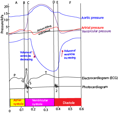
The actions at different points on the graph above are described below:
A Atrium starts to contract: blood flows into the ventricle
B ventricle starts to contract: ventricular pressure exceeds atrial pressure; atrioventricular valve close
C Ventricular pressure exceeds aortic pressure, forcing the aortic valve to open;
blood flows from the ventricle and ventricular volume falls
D Ventricular pressure falls below aortic pressure; aortic valves close
E Ventricular pressure falls below arterial pressure; blood flows from atrium to ventricle; ventricular volume raises rapidly.
F Atrium continuing to fill with blood from the pulmonary vein; atrial pressure exceeds ventricular pressure so blood flows from atrium to ventricle
Cardiac muscle
The most remarkable feature of the heart is the ability to contract rhythmically without fatigue. It owes this property to its muscle known as cardiac muscle. The muscle consists of a network of interconnected muscle fibers.
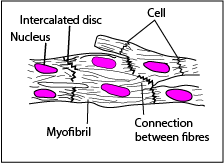
The fibers are divided up into uni-nucleated cells containing myofibrils. The interconnection (intercalated discs) between the fibers ensures a rapid and uniform spread of excitation throughout the walls of the heart which in turn ensures a uniform contraction.
The beating of the heart
Cardiac muscle is myogenic, that is, generate rhythmical contraction from within its muscle. The heart rhythm is initiated by specialized plexus (network)of fine cardiac muscle fibers embedded in the walls of the right atrium close to where the vena cava enters the heart, called the sinoatrial node (SAN) or the pacemaker. The contraction of the heart is preceded by a wave of electrical excitation from SAN and then spread over the heart atria.
When the wave reaches the junction between the atria and ventricle, it excites another specialized group of modified cardiac muscle fibers called the atrioventricular node (AVN), Continuous with the AVN is a strand of modified cardiac muscles fibers called Bundle of Hiss.
The Bundle of Hiss runs down the inter-ventricular septum and fans out over the walls of the ventricle where it breaks up into a network of fibers called Purkinje tissue just beneath the endothelial lining.
When the AVN receives excitation from the atria, it sends impulses down the bundle of Hiss to Purkinje tissue. The impulse then spread out to the cardiac muscle tissue in the walls of the ventricle making it contract.
The role of AVN is to delay electrical impulses from the atria to the ventricle to allow atria contract before the ventricle.
The Purkinje tissue ensures a slow uniform spreading of excitation to the ventricle after the contraction of the atria.
The diagram below is a ventral view of the heart showing the spreading of the electrical excitation that causes contraction.
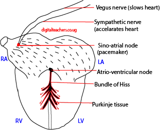
The nerves simply serve to speed up or slow down the heart
Control of contraction
The cardiovascular center in the medulla of the brain controls the rate of heartbeat. Depending on circumstances, the cardiovascular center sends impulse down the sympathetic nerve to the heart, increasing its rate of heartbeat or down the vagus nerves decreasing the rate of heartbeat.
For Revision questions and answers download PDF
Please find free downloadable notes, exams and marking guides of agriculture, biology, Physics, chemistry etc. from digitalteachers.co.ug website.
Dr. Bbosa Science
Thats awesome indeed. I must admit that its a very good job. Thanx so much
Thank you Sir for that job let ALLLAH bless you in it.
Thank you Sir for that job let ALLLAH bless you in it.
It was the best
It is a pleasure to read this weblog, thanks to its up-to-date information and interesting posts. Look into my web page Webemail24 for some really good points and find out more about Network Tunnels.
This is top-notch! I wonder how much effort and time you have spent to come up with these informative posts. Should you be interested in generating more ideas about Information Technology, take a look at my website Seoranko
It appears that you know a lot about this topic. I expect to learn more from your upcoming updates. Of course, you are very much welcomed to my website QN7 about Domains.
dota88
Hello, Neat post. There’s an issue together with your website in internet explorer, might check this?
IE still is the marketplace leader and a large portion of other people will pass over your fantastic writing
because of this problem.
slot demo slot demo slot demo slot demo slot demo
I loved as much as you’ll receive carried out right here. The sketch is tasteful,
your authored material stylish. nonetheless, you command get bought an edginess over
that you wish be delivering the following. unwell unquestionably come more formerly again since exactly the
same nearly very often inside case you shield this increase.
tiktaktogel tiktaktogel tiktaktogel
Hey I know this is off topic but I was wondering if you knew of
any widgets I could add to my blog that automatically tweet my newest twitter
updates. I’ve been looking for a plug-in like this for quite some time and was
hoping maybe you would have some experience with something like this.
Please let me know if you run into anything. I truly enjoy reading your blog and I look forward to your new updates.
targettretinoingelforplanand pharmacy generic
You’ve got a talent for storytelling. Stationary
Thank you ever so for you blog post.Much thanks again. Great.
I enjoy what you guys tend to be up too. This type of clever work and exposure! Keep up the very good works guys I’ve incorporated you guys to my personal blogroll.
I’m amazed, I must say. Seldom do I come across a
blog that’s both educative and interesting, and without a doubt, you have
hit the nail on the head. The issue is something not enough people are speaking intelligently about.
I am very happy that I found this during my hunt for something relating
to this.
Your content is a real gem. 500 ka redeem code
Wow, great article.Thanks Again. Fantastic.
I really enjoy the blog post.Thanks Again. Really Great.
Thanks a lot for the article. Awesome.
Excellent post however I was wondering if you could write a litte more on this subject? I’d be very grateful if you could elaborate a little bit more. Thanks!
Awesome! Its in fact amazing paragraph, I have got much clear idea about from this paragraph.
Please let me know if you’re looking for a writer for your site. You have some really good articles and I think I would be a good asset. If you ever want to take some of the load off, I’d absolutely love to write some material for your blog in exchange for a link back to mine. Please send me an email if interested. Thanks!
My brother recommended I might like this web site. He was totally right. This post truly made my day. You cann’t imagine just how much time I had spent for this information! Thanks!
Woah! I’m really loving the template/theme of this site. It’s simple, yet effective. A lot of times it’s tough to get that “perfect balance” between user friendliness and visual appearance. I must say that you’ve done a excellent job with this. In addition, the blog loads very quick for me on Safari. Exceptional Blog!
With havin so much content do you ever run into any problems of plagorism or copyright violation? My site has a lot of completely unique content I’ve either authored myself or outsourced but it seems a lot of it is popping it up all over the web without my authorization. Do you know any methods to help protect against content from being stolen? I’d definitely appreciate it.
Hi my family member! I want to say that this article is awesome, nice written and include almost all significant infos. I’d like to see more posts like this .
I do like the way you have presented this challenge and it does indeed present me a lot of fodder for thought. Nevertheless, from what I have witnessed, I only wish as the actual feed-back pack on that people today stay on point and not embark on a soap box associated with some other news du jour. All the same, thank you for this exceptional piece and while I can not go along with this in totality, I regard your perspective.
Sweet blog! I found it while surfing around on Yahoo News. Do you have any tips on how to get listed in Yahoo News? I’ve been trying for a while but I never seem to get there! Thank you
Looking forward to reading more. Great article.Thanks Again.
Hi there, just turned into alert to your weblog thru Google, and found that it is really informative. I’m gonna be careful for brussels. I will appreciate if you proceed this in future. Numerous other people might be benefited from your writing. Cheers!
Thank you for the auspicious writeup. It in fact was a amusement account it. Look advanced to more added agreeable from you! However, how could we communicate?
Hmm is anyone else experiencing problems with the images on this blog loading? I’m trying to figure out if its a problem on my end or if it’s the blog. Any feedback would be greatly appreciated.
Admiring the persistence you put into your website and detailed information you present. It’s good to come across a blog every once in a while that isn’t the same unwanted rehashed information. Fantastic read! I’ve bookmarked your site and I’m adding your RSS feeds to my Google account.
ivermectin and pyrantel ivermectin for dogs dosage
Hello, Neat post. There is a problem together with your site in web explorer, could check this… IE still is the marketplace chief and a big element of folks will leave out your magnificent writing due to this problem.
Muchos Gracias for your blog. Cool.
Awesome article.Much thanks again. Great.
Very good information. Lucky me I ran across your blog by accident (stumbleupon).I’ve bookmarked it for later!
Thanks so much for the blog article.Really thank you! Awesome.
I think this is a real great article.Really looking forward to read more. Really Great.
advantages over the other and After that, we started looking
I value the blog. Much obliged.
Great, thanks for sharing this blog post.Really looking forward to read more. Fantastic.
Fine way of describing, and fastidious article to get facts regarding my presentation subject matter, which i am going to present in college.
I appreciate you sharing this post.Really thank you! Really Great.
Really appreciate you sharing this blog post.Much thanks again. Really Great.
Aw, this was a really nice post. Taking the time and actual effort to create a really good articleÖ but what can I sayÖ I hesitate a lot and don’t manage to get nearly anything done.
Awesome article.Really looking forward to read more. Much obliged.
Very good blog post. Will read on…
Amazing issues here. I’m very happy to peer your article.Thank you so much and I am looking ahead to touch you.Will you kindly drop me a e-mail?
Major thanks for the post.Really thank you! Really Cool.
I value the blog post.Really looking forward to read more. Really Great.
You completed some good points there. I did a search on the topic and found nearly all people will agree with your blog.
Hi, you used to write magnificent articles about seattle plumbers, but the last several posts have been kinda lackluster… I miss your tremendous articles. Past several posts are just a little out of track! Seattle Plumbing
Here are the winning numbers for tonight’s Powerball draw.
ed clinics canadian pharmacy – usa pharmacy online
Thanks for the article post.Really looking forward to read more. Will read on…